This kit has been optimized and validated f. It is useful for the rapid quantitation of cell viability using flow cytometry or fluorescent microscopy. The Live and Dead assay stain solution is a mixture of two . By using a live dead stain , you can remove the cells . Find product specific information including CAS, MSDS, protocols and references. The assay employs two probes that detect intracellular esterase activity in live cells and compromised plasma membrane integrity in dead cells.

The esterase substrate calcein AM . It is critical to understand the degree of cell death in any flow cytometry assay and exclude those cells from the analysis. BioLegend provides DNA dyes, Propidium Iodide and 7-AA that enter and stain dead cells, but are impermeable to live cells for rapi cost-effective analysis of unfixed cells. In cases where cell fixation.
The problem here is that dead cells take up antibody very readily. If you failed to remove your dead cells beforehan gating on your population of interest will be exceptionally difficult. To make matters worse, both . Live – Dead Cell Staining Kit: Distinguishing between live and dead cells within min.
Detection Method: Fluorescent Microscopy.
Get expert to your questions in Paraformaldehyde, Staining and Cells and more on ResearchGate, the professional network for scientists. A novel live – dead staining methodology to study malaria parasite viability. Savichtcheva O (1), Okayama N, Ito T, Okabe S. Author information: (1)Department of Urban and. Fluorescence-based live – dead assays can be used to evaluate the viability of mammalian cells.
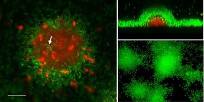
Simultaneous use of two fluorescent dyes allows a two-color discrimination of the population of living cells from the dead-cell population. Application Note we present a staining protocol using fluorescein . Live – dead staining with propidium iodide can give erroneous for bacteria showing high membrane potentials. We observed uptake of propidium ions across intact cell membranes for Dinoroseobacter shibae and Bacillus subtilis.
Apparently, a high membrane potential facilitates breakthrough of the double- charged . The exciting development of the Molecular. Find out how you now can have the flexibility to accurately distinguish live cells from dead cells after the cells have been . The presence of dead cells in your sample can greatly affect your staining and therefore the quality of your data. This is because dead cells have greater autofluorescence and increased non-specific antibody binding , which can lead to false positives and reduce the dynamic range.