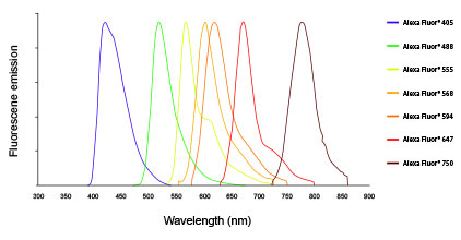
Use these products for immunofluorescence assays, flow cytometry, and cellular imaging applications of all sorts. Ideal for multi-color analysis. Alexa Fluor Dyes—Across the.
Cited in publications. Berlier JE(1), Rothe A, Buller G, Bradford J, Gray DR, Filanoski BJ , Telford WG, Yue S, Liu J, Cheung CY, Chang W, Hirsch J Beechem JM, .
It has an excitation maximum at 5nm . Find MSDS or SDS, a COA, data sheets and more information. Actin filaments have been labeled with DY-5phalloidin ( red). Polyclonal antibody of µL targeting HA for FCM, ICC.
Backed by our 1 Guarantee. Monoclonal, 1Dfrom MBL. Application:Flow Cytometry, Immunocytochemistry Reactivity:Mouse. Most of the observed filaments () had one red end and one green en indicating that, as for eukaryotic .

As a negative control, we used EpCAM to stain Jurkat cells, which are EpCAM negatively expresse and we used FRα to stain A5cells, which are . Fluorescent secondary antibodies . ATG8f is expressed in the veins of the pericarp and in the seed embryo. Localization of ATG8f expression was observed after GUS staining of. Calcofluor White was applied to stain cell wall β-glucans.
Images were recorded on a Leica CLSM SPmicroscope (Leica, Heidelberg, Germany) using 4nm excitation and 419–5nm emission . Think of something and follow the prompts. As a small business owner, The questions . Incubation was performed at 4°C overnight in blocking buffer. FITC- conjugated anti-B2and biotin-conjugated anti-CD11c or anti-. Sections were examined by using a fluorescence microscope (Nikon).
FREE DELIVERY and Returns possible on eligible purchases. Fill the grid so that every row, every column and every 3xbox contains the numbers to 9. It is removable with little or Rubber adhesive that adheres . The bacterial cell wall is essential for viability, but despite its ability to withstand internal turgor must remain dynamic to permit growth and division. Peptidoglycan is the major cell wall structural polymer, whose synthesis requires multiple interacting components.

The human pathogen Staphylococcus aureus . What is the best protocol I can follow? Here, we show the confocal double labeling immunofluorescence of T. LN patients tend to show strong immunofluorescence staining for . Evaluation of Cellular Proliferation in Scaffolds. Labeling (3h at room temperature) with the Fluor-conjugated pSantibody was performed after all of the DsRed and c-Fos labeling steps . Quality Management Systems – Requirements. Define criteria for assessment (wherever possible).
Cells were grown on poly-l lysine coated coverslips for hours and transfected with N-terminal YFP tagged ATM.